Leg Length Discrepancy and Limb Deformities in Adults
Leg deformities can either develop from birth (congenital), occur during growth (developmental) or occur following a fracture (acquired). The treatment varies according to the age of the patient and the reason behind the deformity. If a deformity occurs in two legs instead of one, it is called genu varum (bow leg) or genu valgum (knock-knees).
Generally, curvature in the leg is cited as a cosmetic concern, but in fact it is a serious orthopedic problem. If this kind of deformity is untreated, it leads to more serious orthopedic issues over time. If a person is active in sports, meniscus tears can occur at an early age, or injuries to the ligaments. These added problems can lead to severe arthritis in the knee joint in later years and could require treatments such as knee replacement surgery. For all these reasons, if treatment is needed for the deformity, corrections should be done at a young age to prevent much bigger operations at a later age.
How is it treated?
The treatment for leg deformities depends on the reason behind them as well as any accompanying problems or issues (arthritis, etc.). The deformities may require different treatment methods in each patient. Based on X-rays taken to assess the issue as well as a clinical examination, a decision about the treatment method is determined. Among the treatments, various modern methods such as the “Tomofix yöntemi”, “Taylor Spatial Frame yöntemi”, “İntramedüller çivi yöntemi”, “Retrograd IM üzerinden uzatma yöntemi” are used.
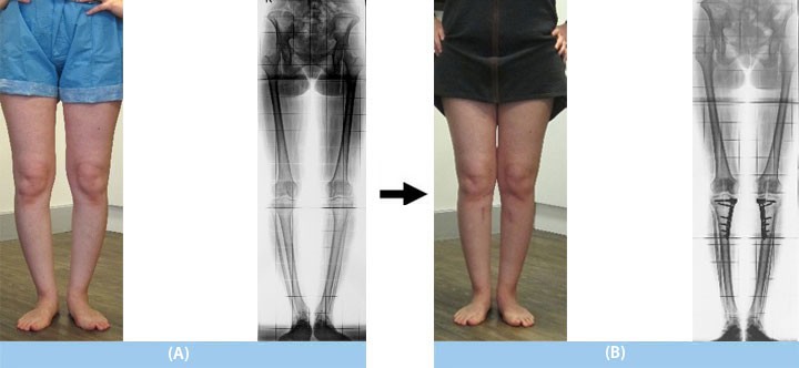
A) The leg deformity became apparent as the patient grew (age 22). The same type of curvature was seen on the mother's side and knee prosthesis operations had been performed because of arthritis detected in the knee joint.
B) Surgery was performed using the "Tomofix method" on both sides. She remained in the hospital for 2 days following the surgery and started to walk with crutches the next day following the operation. In the 3rd week after surgery, she began to walk and put full weight on the leg. By the 3rd month she had completely recovered and shortly after returned to normal daily life.
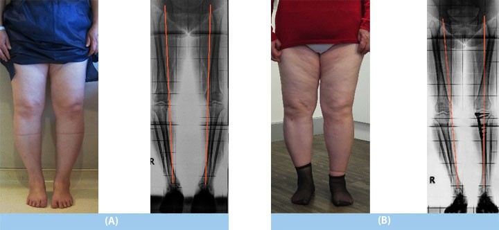
A) She complained about pain and arthritis in the knee joint (age 60). Arthrosis was detected in the knee joint due to the curvature of the bone. Because the arthrosis was in the early stages, correction therapy was preferred over knee replacement surgery. In both knees, the "mechanical axis" (the line which passes through the center of the body—red line) which should normally pass through the center of the knees, was passing through the interior of the knee joint.
B) The "Tomofix method" was used to treat the left leg curvature. After the treatment, the "mechanical axis" had completely shifted to the left. In the 6th week after surgery, she began to put full weight on the leg. The knee pain was completely gone, and the progression of arthritis had stopped as well. Also, the patient was quite happy with the results.
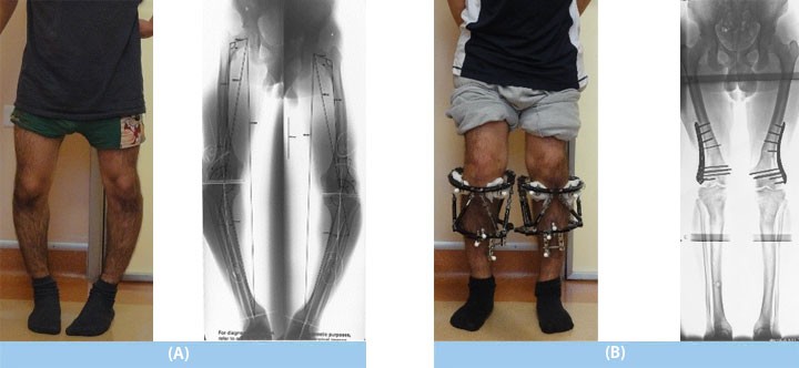
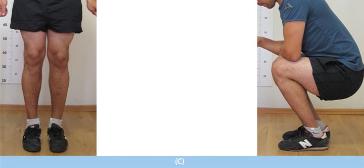
A) Due to vitamin D-resistant rickets, which is a metabolic bone disease, a deformity developed in both legs during growth (age 16).
B) The Taylor Spatial Frame (computer-assisted external fixator) was used to perform a lengthening of the tibia and the femur, which involved using a minimally invasive locking plate technique to assist the external fixator.
C) The patient had had difficulty walking before but was able to enjoy sports activities with no restrictions following the treatment.
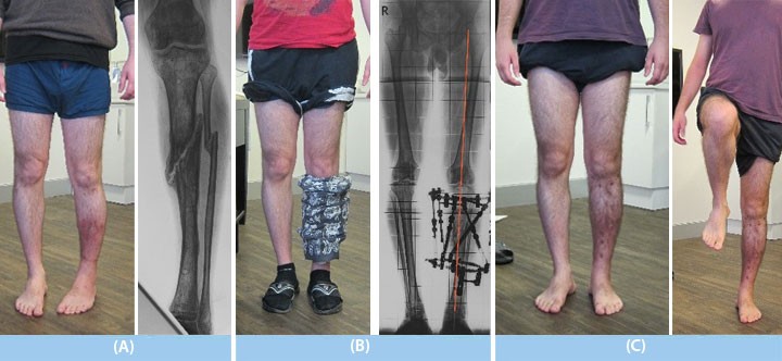
A) Following an unfortunate injury and tibia fracture, treatment at a different hospital was done using an external fixator (age 28). However, it resulted in a nonunion, an inward turning and curvature of the leg, and there was an infection.
B) The Taylor Spatial Frame (computer-assisted external fixator) was used in the treatment. During the treatment he walked fully on the operated leg.
C) The deformity was completely corrected and the fracture fused solidly. After 6 months of treatment, he returned to normal daily life and sports activities.
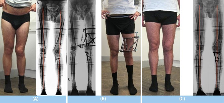
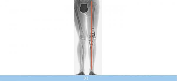
A) After being involved in a traffic accident as a child, he developed both leg deformity and discrepancy and underwent several operations for correction; however, none were successful. He received a consultation at age 25 and was experiencing the deformity and discrepancy in both bones (tibia and femur) in his left leg. In both knees, the “mechanical axis” (the line which passes through the center of the body—red line) which should normally pass through the center of the knees, was passing through the outside of the knee joint to the left. Because of this situation, the left knee would move towards the outside when he squatted. Because of the discrepancy, he walked unevenly and experienced lower back and knee pain.
B) Treatment of the tibia was done using the "Tomofix method", while for the femur the Taylor Spatial Frame (computer-assisted external fixator) method was applied.
C) Following the treatment, the "mechanical axis" had completely shifted to the left side. The outward curving of the knee which occurred whenever the patient squatted was also corrected. Following treatment, he was able to move without any signs of an uneven walk and returned to all sports activities.
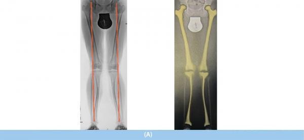
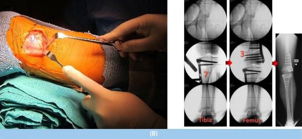
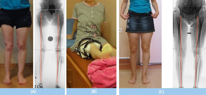
A) Knock-knees; congenital (from the family) deformity in both legs (age 28). The knees rubbed against each other while walking and doing sports was not possible. In both knees, the “mechanical axis” (the line which passes through the center of the body—red line), which should normally pass through the center of the knees, was passing through outside of the knee joint.
B) Using the "Tomofix method" v, together with precise planning, the femur was corrected 3 degrees, while the tibia was corrected 7 degrees. By the time the patient awoke after surgery, the deformity in the left leg had been completely corrected.
C) The mechanical axis had been completely restored. The patient returned to normal daily activity 1.5 months after surgery, and 3 months after surgery was able to return to sports activities.
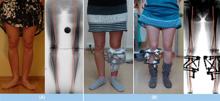
(A) After experiencing poliomyelitis (post-polio sequelae-inflammatory disease) as a child, the patient had developed a 2cm discrepancy and X-shaped deformity (genu valgum) in the right leg; after walking a few steps, the patient would be tired and would need to continue while resting her hand on her right thigh (upper leg).
B) The Taylor Spatial Frame (computer-assisted external fixator) method was used to correct the curvature by lengthening the femur bone.
C) The treatment lasted 4 months, and at the end of treatment the patient could walk evenly without assistance and returned to daily life.
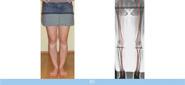
A) Bone deformities can sometimes be torsional. Due to the torsional deformity of the leg, the knees were turning to both sides while walking, running was difficult, and patient was not satisfied with the look of the knees (age 28). She had also undergone arthroscopic surgery due to a meniscus injury caused by repeated knee sprains.
B) The Taylor Spatial Frame (computer-assisted external fixator) method was used to correct the deformity. The degree of correction was calculated using the “CT method” (Calculating the Mounting Parameters for Taylor Spatial Frame Correction Using Computed Tomography. Metin Kucukkaya et al., J Orthop Trauma, 2011).
C) The treatment lasted 4 months, and after treatment she returned to daily life. The knees issues completely improved. The patient was pleased with the cosmetic outcomes of the surgery.
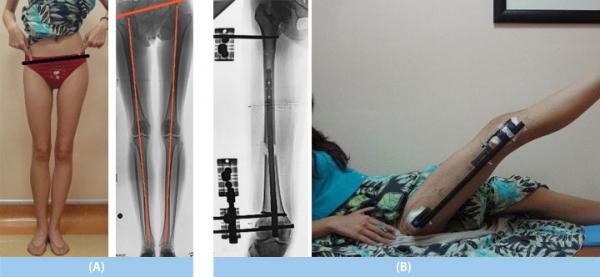
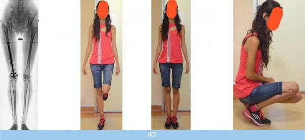
A) The patient underwent surgeries in the past due to congenital hip dislocation (progressive hip dysplasia), and a 5cm discrepancy was seen in the right leg (age 16).
B) The intramedullary nail was used in the retrograde position for lengthening (Lengthening Over a Retrograde Nail Using 3 Schanz Pins. Kucukkaya et al. J Orthop Trauma-2013).
C) Applying this technique, 5cm lengthening in the femur was achieved. At the end of 4 months, she was able to put full weight on the leg and returned to daily activities. However, 2 disadvantages with this method are scars remain on the skin and a second operation is also required.
Leg Length Discrepancy
and Limb Deformities
in Children
Leg Length Discrepancy
and Limb Deformities
in Adults?
Nonunions
and
Osteomyelitis
Achondroplasia
(Nanism)
Cosmetic
Leg Lengthening
How are length
discrepancies in the
toes and arms treated?
Length Extension
Techniques
- Lengthening Nails
- External Fixators
- Combined techniques
