How are leg length discrepancies and limb deformities in children treated?
The aim of treatment is to achieve results that erase all memory of the child's leg discrepancy or deformity in adulthood. In children, the treatment depends on the cause as well as any problems associated with the condition. The treatment required for limb deformities may be different in each patient. Based on X-rays taken to assess the situation as well as a clinicial examination, a decision about the treatment is determined. Among the treatments available, the "Precice magnetic rod (lengthening device)", "Taylor Spatial Frame method", "Intramedullary nail method", "8-Plate (guided growth) method" are some of the various modern methods which are used.
An important point about lengthening procedures done for children: The rate of complication is very high in lengthening surgery performed before the age of 5 (growth plate suppression, infection, new deformities appearing during the lengthening, etc.). For the first lengthening surgery, the child should have reached the age of 5.
An important note about lengthening for children with achondroplasia: In children with achondroplasia, 2 bones on the same side (upper thigh and lower tibia) should not be lengthened. Although treatment like this was done in the past, many doctors have given it up due to objections being raised about it. It is believed that the natural growth potential of the child would be suppressed or inhibited because performing a lengthening on the same side suppresses the growth plate.
Precice Magnetic Rod Method
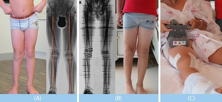
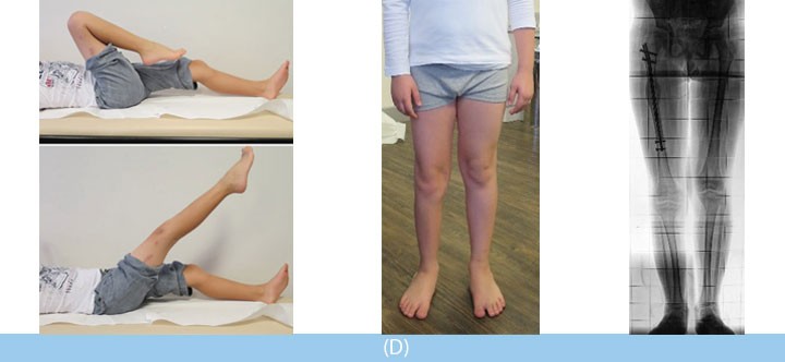
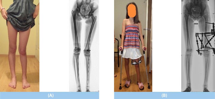
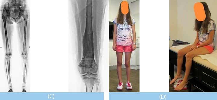
"Fibular hemimelia" is a condition characterized by limb length discrepancy, outward curvature and difficulty shows up at around the age of 8. The aim of treatment is to achieve a completely normal leg with no discrepancy or deformity by the time the child reaches ages 14-16, and to deliver a treatment with minimally invasive techniques, e.g. using the least amount of operative devices and placing devices internally without the use of an external fixator. In order to do this, a plan of treatment was made.
A) The first action was delayed for 2 years, until the right age, and arch supports were placed inside the shoes.
B) Based on the result of the calculations, the deformity was corrected using the"8-Plate" (guided growth) technique (similar to dental restoration techniques applied to the teeth). In this method, a 2cm incision was made to place the 8-Plate in the growth plate area, and the patient needed to remain in the hospital for one day following the procedure. The following day, all daily activities were allowed, including sports. 3 months later, after it was determined that the mechanical axis (corresponding to the curvature) had been restored with X-rays, the second stage of treatment was started.
C) In the second stage, the 8-Plate was removed and an intramedullary instrument (Precice-magnetic rod) was put in place for the lengthening. After the surgery, a 5cm lengthening was achieved through an external remote during a 2-month period, and the patient was allowed to walk with crutches.
D) In the 4th month after surgery, the patient was walking with no assistance and returned to normal daily activities. The treatment was planned so that the child did not lose any time in school.
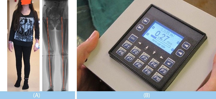
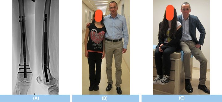
A) The patient arrived to us at age 12 experiencing a 4cm discrepancy in the right femur and a deformity had developed towards the outside. She had undergone surgery before to address issues with her spine and hip.
B) To address the leg deformity, an "8-Plate" an intramedullary lengthening device (Precice magnetic rod) was positioned, and at the same time the leg deformity was corrected; an overall lengthening of 4 cm was achieved through a 1mm daily lengthening done by controlling the tool from the outisde. She walked with crutches during the lengthening period,
C) but in the 4th month after the surgery she began to put full weight on the leg without the assistance of crutches.
8-Plate Guided Growth Method
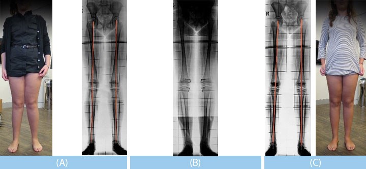
A) A crooked knee (deformity in both knees), walking difficulty (knees rubbing together) and outward curving of the toes (Hallux valgus) was seen (age 11). She had been treated in the past for the Hallux valgus and surgery had been suggested. In reality, the Hallux valgus in the patient is caused by the knee deformity and does not require a separate treatment.
B) The 8-Plate (guided growth) technique (similar to dental restoration techniques applied to the teeth) was used to correct the deformity within 4 months. In this method, a 2cm incision was made to place the 8-Plate in the growth plate area, and the patient needed to remain in the hospital for one day following the procedure. The following day, all daily activities were allowed without any restriction, and sports activities continued.
C) At the end of treatment (4 months later), the deformity in both knees was corrected and any walking difficulty was resolved.
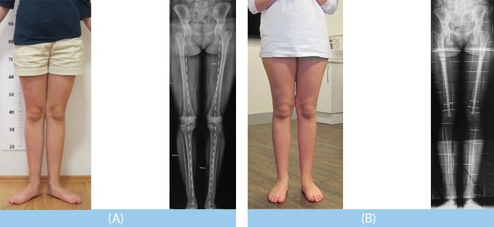
Bone deformities can sometimes be torsional.
A) Because of the torsional leg deformity, the knees would turn in both directions (inward and outward) while walking, the patient experienced trouble running, and both the patient as well as the family were unhappy with the overall appearance (age 17).
B) The intramedullary nail technique was used in the treatment. The largest surgical incision was 1.5 cm, and 6 stitches were applied during surgery. After the surgery, the patient was able to perform sports activities much better.
A) The congenital limb discrepancy and outward curvature of the left leg became apparent by the age of 8. The discrepancy could not be compensated for with shoes lifts.
B)The extent of the discrepancy that would develop through growth by the ages of 14-16 was calculated and a lengthening of the femur was planned. The Taylor Spatial Frame (computer-assisted external fixator) was used in the lengthening to correct the femur deformity by 8.5 cm.
C) The treatment lasted 5.5 months. At the end of treatment, she began to walk with no discomfort, experienced no restriction in movement of the joints, and soon returned to daily life and sports activities.
D) Because the operation was done at the correct time (appropriate age) and accurate calculations were done, by the time the patient reached age 15, the predicted outcomes were achieved and the legs were equal length.
Taylor Spatial Frame Method
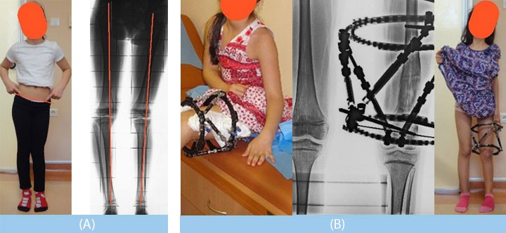
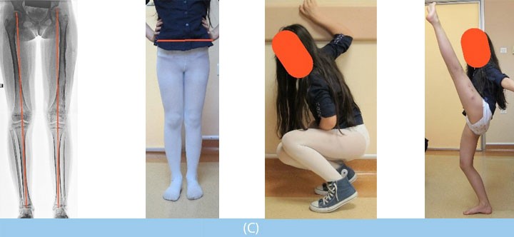
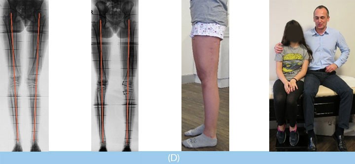
A) The congenital discrepancy and outward curvature on the left leg became apparent by the age of 8. The discrepancy could not be compensated for with shoes lifts. The "mechanical axis", which is the line that normally passes through the middle of the knee joint, appears to pass from the outside of the knee joint in the left leg (red line).
B) The extent of the discrepancy that would develop through growth by the ages of 14-16 was calculated and a lengthening of the femur was planned. The Taylor Spatial Frame (computer-assisted external fixator) method was used in the lengthening to correct the femur deformity by 8 cm and at the same time correct all the deformity. The treatment lasted 5.5 months. In order to calculate the discrepancy, it was very important that the surgery be done at the correct time (appropriate age).
C) At the end of treatment, she began to walk with no discomfort, experienced no restriction in movement of the joints, and soon returned to daily life and sports activities.
D) When she reached the age of 12, there was a slight deviation of the "mechanical axis" (red line) in the left leg. Because the operation had been done at the correct time (appropriate age) and the correct calculations had been done, discrepancy had not occurred. Finally, the 8-plate (guided growth) technique (similar to dental restoration techniques applied to the teeth) restored the "mechanical axis". In this session, surgical scars remaining from the previous surgery were corrected by the plastic surgery team and the treatment was ended in a way whereby no other problems would appear later in life.
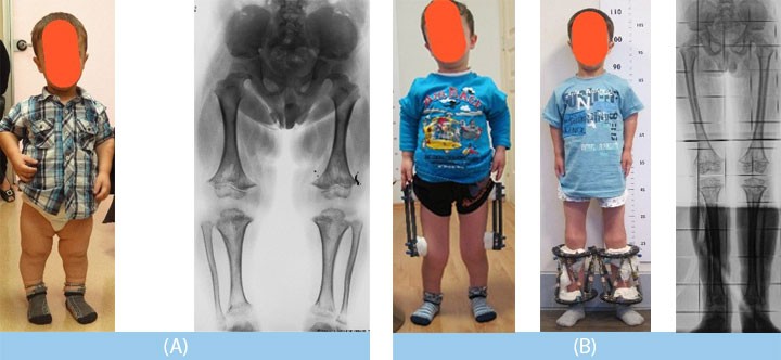
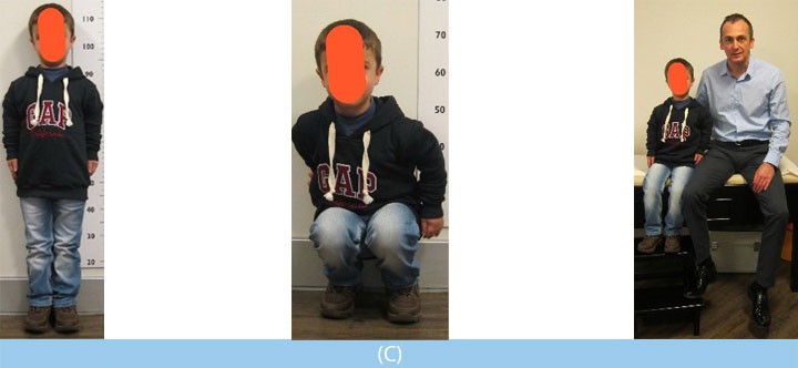
A) The short stature of the patient had been caused by achondroplasia. If left untreated, it was calculated that the patient would reach a maximum height of 90 cm by the time of adulthood.
B) The treatment for the achondroplasia was begun at the age of 5, the ideal age of a first lengthening, and both femurs were lengthened by 12 cm. A year later at the age of 6.5, the tibiae were lengthened 11 cm.
C) In this way, by the time the patient reached the age of 7, a total lengthening of 23 cm had been done.
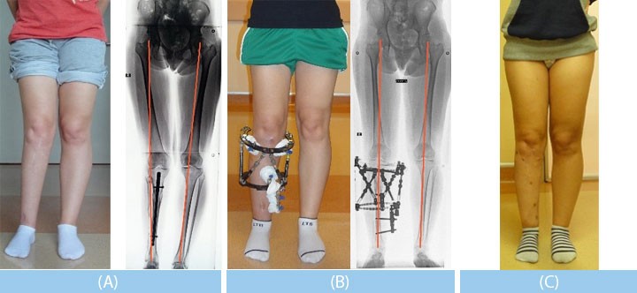
A) Deformity and discrepancy in the right leg had developed during growth due to a spinal disease. The right leg had been lengthened before; however, both problems reappeared (age 16).
B) The Taylor Spatial Frame (computer-assisted external fixator) method was used to correct the tibia curvature as well as do the lengthening at the same time.
C) The treatment lasted 3 months, and the patient began to walk without discomfort at the end of treatment and returned to daily life.
Leg Length Discrepancy
and Limb Deformities
in Children
Leg Length Discrepancy
and Limb Deformities
in Adults?
Nonunions
and
Osteomyelitis
Achondroplasia
(Nanism)
Cosmetic
Leg Lengthening
How are length
discrepancies in the
toes and arms treated?
Length Extension
Techniques
- Lengthening Nails
- External Fixators
- Combined techniques
